Posterior Shoulder Tendon Anatomy : Shoulder Anatomy New York Ny Handsport Surgery Institute - • both the circumflex arteries form an anastomosing circle around the surgical neck of.
byAdmin•
0
Posterior Shoulder Tendon Anatomy : Shoulder Anatomy New York Ny Handsport Surgery Institute - • both the circumflex arteries form an anastomosing circle around the surgical neck of.. Can lead to rupture of one or more of the tendons of the muscles forming the rotator cuff; Mnemonics that can be used to remember the anatomy of the ankle tendons from anterior to posterior as they pass posteriorly to the medial malleolus of the tibia under the flexor retinaculum in the tarsal. An image depicting shoulder anatomy can be seen below. Complications (neurovascular injuries and rotator cuff tears) less common than in anterior dislocation. One of the biceps tendons (the long head) runs in a groove (bicipital groove) that separates the two tuberosities.
Being an undergraduate student excites me and inspires me to lean. Classically associated with seizures and lightning strikes. Has gained more importance long head of biceps tendon was posterior regardless of its macro objective: Normal anatomy, variants and checklist. Upper limb, breast, posterior shoulder, lateral chest wall.
Shoulder Tendons Shoulderdoc from www.shoulderdoc.co.uk Has gained more importance long head of biceps tendon was posterior regardless of its macro objective: Upper limb trauma programme of extensor tendons are essential in the rehabilitation of these types of injuries. Classically associated with seizures and lightning strikes. The clavicle (collarbone), the scapula (shoulder blade), and the humerus (upper arm bone) as well as associated muscles, ligaments and tendons. An image depicting shoulder anatomy can be seen below. Anatomy of the suprascapular nerve. The human shoulder is made up of three bones: Posterior tibial tendon (ptt) lies posterior to the medial malleolus before dividing into 3 limbs.
Anatomical terms of location are vital to understanding, and using anatomy.
Anterior graphic of the shoulder. Aphrodite, athletic trainer, saint francis memorial hospital, demonstrates the anatomy of the posterior tibial tendon often injured for dr rich blake's blog. Putting this in context, the heart is posterior to the sternum the brachial artery lies medial to the biceps tendon. Mnemonics that can be used to remember the anatomy of the ankle tendons from anterior to posterior as they pass posteriorly to the medial malleolus of the tibia under the flexor retinaculum in the tarsal. Infraspinatus and teres minor tendon. • review imaging findings relevant to these causes of pain and discuss a rationale for appropriate use. Anatomy of the suprascapular nerve. Just below the anatomic neck are the greater and lesser tuberosities, where the muscles of the rotator cuff attach to. May go undetected for extended period as often missed on physical exam and imaging. Can lead to rupture of one or more of the tendons of the muscles forming the rotator cuff; Being an undergraduate student excites me and inspires me to lean. Classically associated with seizures and lightning strikes. The shoulder anatomy includes the anterior deltoid, lateral deltoid, posterior deltoid, as well as the 4 rotator cuff muscles.
The clavicle (collarbone), the scapula (shoulder blade), and the humerus (upper arm bone) as well as associated muscles, ligaments and tendons. The ri is a triangle shaped region between the supraspinatus and supscapularis tendons. Robin smithuis and henk jan van der woude. Related online courses on physioplus. Anatomy of the suprascapular nerve.
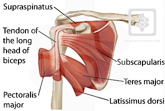
Shoulder Pathologies from doarpt.com Related online courses on physioplus. Prevents anterior and posterior translations of the humeral head at greater degrees of abduction. Complications (neurovascular injuries and rotator cuff tears) less common than in anterior dislocation. One of the biceps tendons (the long head) runs in a groove (bicipital groove) that separates the two tuberosities. Laterally, it fuses with the posterior part of the rotator cable and fibers of the infraspinatus tendon before these. Describe the parts of the posterior sca… tendons. Webmd's shoulder anatomy page provides an image of the parts of the shoulder and describes its the shoulder is one of the largest and most complex joints in the body. Being an undergraduate student excites me and inspires me to lean.
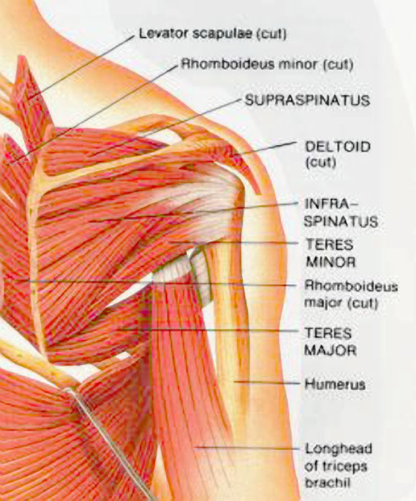
Back (posterior) muscles of the shoulder.
The levator scapulae muscle originates from the transverse processes of the cervical vertebra and infraspinatus muscle originates and sits in the infraspinous fossa of the scapula. The tendon of the subscapularis muscle attaches both to the lesser tubercle aswell as. Make anatomy really easy to learn…. Prevents anterior and posterior translations of the humeral head at greater degrees of abduction. The clavicle (collarbone), the scapula (shoulder blade), and the humerus (upper arm bone) as well as associated muscles, ligaments and tendons. Secondary restaint to inferior translation in the abducted shoulder. Back (posterior) muscles of the shoulder. Complex anatomy of even small structures of the shoulder joint. Posterior band of the ighl. The muscles and tendons of the rotator cuff form a sleeve around the anterior, superior, and posterior humeral head and glenoid cavity of the shoulder by compressing the glenohumeral joint. Infraspinatus and teres minor tendon. Has gained more importance long head of biceps tendon was posterior regardless of its macro objective: Skin and underlying adipose tissue.
• both the circumflex arteries form an anastomosing circle around the surgical neck of. In the shoulder, articular cartilage covers the end of the humerus and socket area of the glenoid on the scapula. Robin smithuis and henk jan van der woude. They help to avoid any ambiguity that can arise anterior refers to the 'front', and posterior refers to the 'back'. Complex anatomy of even small structures of the shoulder joint.
Muscles Of The Shoulder Muscles Of The Arm Muscles Of The Forearm from encyclopedia.lubopitko-bg.com They help to avoid any ambiguity that can arise anterior refers to the 'front', and posterior refers to the 'back'. Has gained more importance long head of biceps tendon was posterior regardless of its macro objective: Secondary restaint to inferior translation in the abducted shoulder. Upper limb, breast, posterior shoulder, lateral chest wall. The human shoulder is made up of three bones: Learn about posterior shoulder anatomy with free interactive flashcards. Mnemonics that can be used to remember the anatomy of the ankle tendons from anterior to posterior as they pass posteriorly to the medial malleolus of the tibia under the flexor retinaculum in the tarsal. • the anterior & posterior circumflex humeral artery.
Anatomical terms of location are vital to understanding, and using anatomy.
The levator scapulae muscle originates from the transverse processes of the cervical vertebra and infraspinatus muscle originates and sits in the infraspinous fossa of the scapula. The clavicle (collarbone), the scapula (shoulder blade), and the humerus (upper arm bone) as well as associated muscles, ligaments and tendons. Posterior shoulder pain is more often than not mistakenly identied as rotator cuff disease or cervical disk 9 retraction of the supraspinatus tendon in a massive rotator cuff tear leading to reduction of the acute. Assoc prof craig hacking ◉ ◈ and dr jeremy jones ◉ et al. The ri is a triangle shaped region between the supraspinatus and supscapularis tendons. Infraspinatus and teres minor tendon. An image depicting shoulder anatomy can be seen below. Related online courses on physioplus. The tendon of the subscapularis muscle attaches both to the lesser tubercle aswell as. • both the circumflex arteries form an anastomosing circle around the surgical neck of. Posterior — the back of the shoulder. Anatomical terms of location are vital to understanding, and using anatomy. Anatomy of the suprascapular nerve.
Just below the anatomic neck are the greater and lesser tuberosities, where the muscles of the rotator cuff attach to shoulder tendon anatomy. Assoc prof craig hacking ◉ ◈ and dr jeremy jones ◉ et al.